Vitreous Humor Eye Diagram

Eye Facts Cool Kid Facts

Human Eye Definition Structure Function Britannica

Associated Retina Consultants Diagram Of The Eye Phoenix
.jpg)
Structure And Function Of The Eyes Eye Disorders Merck Manuals

Eye Microdissection Iris Pharma

Human Eye Anatomy Parts Of The Eye Explained Eye Anatomy
The vitreous chamber is the largest cavity in the eye which makes the vitreous humour the most prominent liquid in the eye.
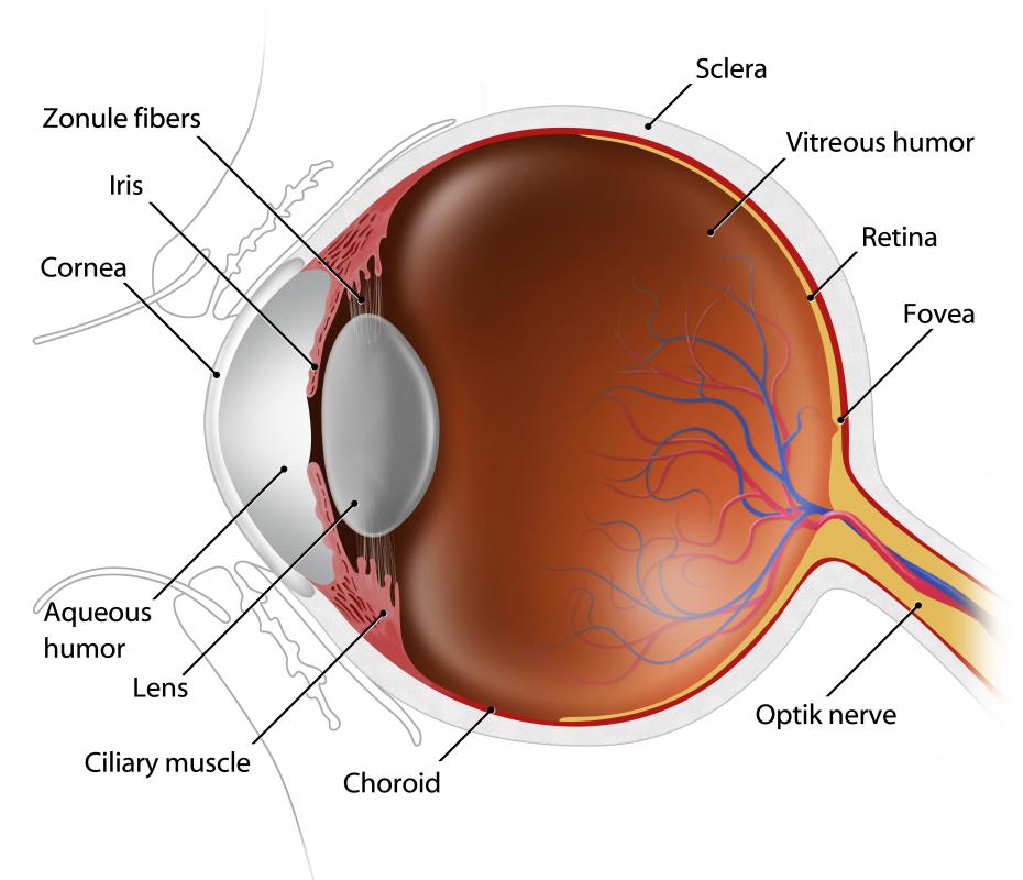
Vitreous humor eye diagram. The vitreous humor is a transparent colorless gelatinous mass that fills the space in the eye between the lens and the retina it is surrounded by a layer of collagen called the vitreous membrane or hyaloid membrane or vitreous cortex separating it from the rest of the eye. It makes up four fifths of the volume of the eyeball. The anterior chamber the vitreous chamber and the posterior chamber. The vitreous humor also known simply as the vitreous is a clear colorless liquid that.
We are pleased to provide you with the picture named vitreous humor in human eye we hope this picture vitreous humor in human eye can help you study and research. Present at birth it changes little until we enter our fifth decade when it begins to shrink which can lead to disorders ranging from harmless floaters to a vision impairing retinal detachment. It is the largest of the cameras and occupies about 80 of the eye. As we age vitreous humor shrinks.
These substances are called the vitreous humor and aqueous humor. Status of vitreous humour over time. It is a clear gel like substance that occupies the space behind the lens and in front of the retina at the back of the eye. The vitreous humour is fluid like near the centre.
Diagram of the vitreous cavity during posterior vitreous detachment. Anatomy in the most basic of the anatomical terms the eye is divided into three sections. The vitreous camera is placed on the back of the eyeball. Nov 28 2018 explore khobbsgreen s board posterior vitreous detachment followed by 312 people on pinterest.
Filling most part of the eyes vitreous humour helps maintain the shape of the eyes intact. Vitreous humor thus performs the important function of enabling eyesight in a clear manner. The vitreous cavity lies between the lens and the back of the eye. Over the next 1 to 3 months the vitreous gel further condenses and the sides of the gel also separate from the retina until the pvd is complete and the vitreous gel is attached to the retina only at the vitreous base see figure 1.
It is through this liquid that the light rays pass through the eye lens to the retina. See more ideas about posterior vitreous detachment detachment eye health. Vitreous is a. For more anatomy content please follow us and visit our website.
Light that is focused into the eye by the cornea and lens passes through the vitreous onto the retina the light sensitive tissue lining the back.

Eye Pain Symptoms Signs Causes Treatment

Understanding Aqueous Humor And Vitreous Humor The Differences
What Is The Vitreous Humor With Pictures

Human Eye Anatomy Parts Of The Eye Explained Eye Anatomy

Vitrectomy For Floaters The American Society Of Retina Specialists

Underwater Vision In Penguins Cormorants And Sea Gypsies

Human Eye Anatomy How The Human Eye Works

The Human Eye Is Wired Backwards And These Scientists Think

2 5 The Eye University Physics Volume 3 Openstax

Cross Section Through The Human Eye Diagram By The Author

Understanding Aqueous Humor And Vitreous Humor The Differences

Human Eye Diagram Images Stock Photos Vectors Shutterstock

About My Eyes Optical Express Optical Center

Internal Anatomy Of The Eye Diagram Quizlet

Structure And Function Of The Eyes Eye Disorders Merck Manuals

Eye Anatomy

File Eye Diagram No Circles Border Svg Wikipedia

Anatomy Of The Eye Human Eye Anatomy Owlcation

Eye Anatomy

Understanding Aqueous Humor And Vitreous Humor The Differences

How The Human Eye Works Cornea Layers Role Light Rays

File Schematic Diagram Of The Human Eye Png Wikipedia

File Eye Diagram Without Text Gif Wikimedia Commons

Understanding Aqueous Humor And Vitreous Humor The Differences

Definition Of Vitreous Humor Nci Dictionary Of Cancer Terms

Human Eye Anatomy Parts Of The Eye Explained Eye Anatomy

Human Eye Diagram Images Stock Photos Vectors Shutterstock
Normal Vision

How The Eye Works Norfolk Virginia Beach Va

Definition Of Vitreous Humor Nci Dictionary Of Cancer Terms

2 5 The Eye University Physics Volume 3 Openstax

Associated Retina Consultants Diagram Of The Eye Phoenix

Movement And Structure Of The Eye

Diabetic Eye Disease Non Proliferative Diabetic Retinopathy
Vitreous Body Wikipedia

Our Eyes Work Like Camera S Discovery Eye Foundation

Understanding The Different Parts Of Your Eye All About Eyes

Human Eye Definition Structure Function Britannica

Structure And Function Of The Human Eye

Eye Facts Cool Kid Facts

How The Human Eye Works Cornea Layers Role Light Rays

Parts Of The Eye

Optometry Humor Vitreous Humor J Aqueous Humor K Retina

Eye Pain Symptoms Signs Causes Treatment

Definition Of Vitreous Humor Nci Dictionary Of Cancer Terms
Eyeball Diagram Of Membranes That Surround Transparent

Eye Pain Symptoms Signs Causes Treatment

Https Www Sps186 Org Downloads Basic 343602 Sheep 20eye 20dissection Pdf

Ultraviolet Radiation Oxidative Stress Affects Eye Health Ivanov

Ciliary Body Images Stock Photos Vectors Shutterstock

Eye Vitreous Aqueous Humor Diagram Function Body Maps

Human Eye Anatomy How The Human Eye Works

Eye Anatomy Ocular Anatomy Vision Conditions Problems

Human Eye Anatomy How The Human Eye Works

Eye Anatomy Detail Picture Image On Medicinenet Com

Chapter 28 Concept 28 5

Eye Anatomy Ocular Anatomy Vision Conditions Problems

What Is The Vitreous Humor With Pictures

Parts Of The Eye

Normal Vision

Vitreous Body Wikipedia

Humor Vitreous Humor Eye Diagram Wiring Diagram Images

Eye Diagram Free Vector Art 142 Free Downloads

Cow S Eye Dissection Eye Diagram

Posterior Vitreous Detachment The American Society Of Retina

Parts Of The Eye Their Function Robertson Optical And Optometry

How Vision Works Eye Science Projects Experiments Hst

Understanding Aqueous Humor And Vitreous Humor The Differences

Ultraviolet Radiation Oxidative Stress Affects Eye Health Ivanov

Your Eyes Johns Hopkins All Children S Hospital
/GettyImages-695204442-b9320f82932c49bcac765167b95f4af6.jpg)
Eyes Johns Hopkins All Children S Hospital

Parts Of The Eye American Academy Of Ophthalmology

Human Eye Definition Structure Function Britannica

Your Eyes For Kids Nemours Kidshealth

Human Eye Diagram Images Stock Photos Vectors Shutterstock

Movement And Structure Of The Eye

Https Www Sps186 Org Downloads Basic 343602 Sheep 20eye 20dissection Pdf

Vitreous Body Wikipedia

Eyeball Diagram Of Membranes That Surround Transparent

Eyes Anatomy Overview Parts And Functions Biology Dictionary

Parts Of The Eye American Academy Of Ophthalmology

Parts Of The Eye Their Function Robertson Optical And Optometry

Figure Figure 1 Anatomy Of The Pdq Cancer Information

Anatomy Of The Eye Human Eye Anatomy Owlcation

Labeled Eye Diagram Science Trends

Eyes Johns Hopkins All Children S Hospital

Parts Of The Eye American Academy Of Ophthalmology

Human Eye Anatomy Parts Of The Eye Explained Eye Anatomy

Anatomy Of The Eye Red Rover Ventures

2 5 The Eye University Physics Volume 3 Openstax

Blood Vessels And Nerves Of The Eye Anatomy Kenhub

Your Eyes Johns Hopkins All Children S Hospital
